North India Statesman
When an unexpected injury strikes, persistent pain refuses to fade, or a physician orders an urgent medical scan, patients often find themselves facing a confusing choice of imaging modalities: an X-ray, a CT scan, or an MRI. While all three peer deep inside the human body without an incision, they rely on entirely distinct physical principles, carry different safety profiles, and serve specific diagnostic purposes. Deciding which scan is necessary is not a matter of choosing the “most advanced” test, but matching the exact anatomical question with the appropriate diagnostic technology.
The Core Answer: How the Three Scans Differ at a Glance
Medical imaging rests on three separate pillars:
-
Conventional X-rays send a brief burst of electromagnetic radiation through the body onto a digital detector. Dense structures like cortical bone absorb the rays and appear bright white, while air-filled cavities like the lungs appear black. It delivers a fast, flat, two-dimensional picture.
-
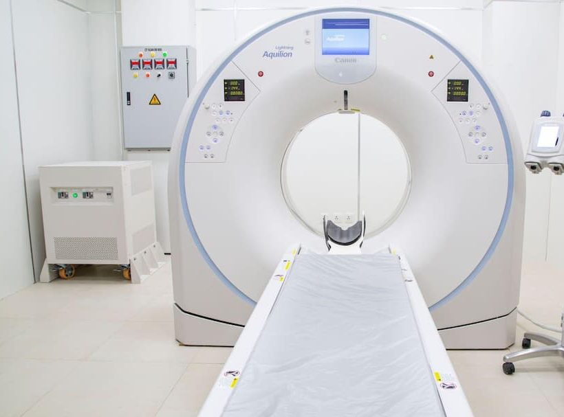
Computed Tomography (CT) Scans also deploy X-rays, but instead of taking a single static snapshot, a motorized X-ray tube rotates rapidly around the patient inside a circular gantry. Powerful computer algorithms piece together thousands of thin cross-sectional “slices,” assembling intricate 3D representations of internal organs, blood vessels, and bones.
-
Magnetic Resonance Imaging (MRI) abandons ionizing radiation altogether. Instead, it utilizes an intense magnetic field—typically 1.5 to 3.0 Tesla—combined with pulsed radiofrequency waves to excite hydrogen atoms within bodily tissues. As these protons realign, they emit distinct radio signals picked up by specialized receiver coils, generating peerless, high-resolution cross-sectional maps of soft tissues, nerve pathways, and ligaments.
Understanding Conventional X-rays: The Frontline Workhorse
Conventional radiography remains the most widely accessible and rapid imaging tool across primary healthcare centres, urgent care setups, and district hospitals. Because bone tissue contains high concentrations of calcium—a high atomic number element—it attenuates X-ray beams far more effectively than adjacent muscular or adipose tissue.
Doctors order an X-ray when they suspect traumatic bone fractures, joint dislocations, severe dental decay, arthritis progression, or foreign body ingestion. In cardiopulmonary medicine, a standard chest X-ray (CXR) serves as the frontline evaluation for bacterial pneumonia, fluid accumulation in the pleural cavity, pneumothorax, or cardiomegaly (enlarged heart).
The primary clinical advantage of an X-ray is sheer speed and convenience. The procedure takes mere seconds, requires no physical confinement, and costs relatively little—often between ₹250 and ₹800 at local diagnostic laboratories. The radiation dose is minimal; an ordinary chest X-ray exposes a patient to roughly 0.1 mSv, equivalent to about 10 days of natural ambient background radiation. However, its major limitation is two-dimensional overlap: overlapping tissue shadows can easily obscure subtle microfractures, early-stage tumours, soft-tissue tears, and internal abdominal bleeds.
Computed Tomography (CT): Precision in Critical Emergencies
When doctors require cross-sectional depth, volumetric measurement, or immediate insight during acute trauma, the multi-detector CT scan is the standard of care. Because a modern multi-slice scanner completes full rotational acquisitions in fractions of a second, an entire chest, abdomen, or pelvis can be thoroughly surveyed in less than a minute.
In emergency medicine, speed saves brain tissue and organ function. A non-contrast head CT scan is the universal protocol for acute stroke triage, instantly differentiating between an ischemic infarction and a life-threatening intracerebral haemorrhage before clot-busting thrombolytic drugs are administered. Similarly, in high-speed vehicular collisions or blunt abdominal trauma, contrast-enhanced CT scans immediately detect lacerations in the liver or spleen, aortic dissections, and complex pelvic fractures that standard X-rays miss. In pulmonology, high-resolution computed tomography (HRCT) remains the benchmark for evaluating interstitial lung diseases, pulmonary fibrosis, and pulmonary embolisms.
CT scanning carries two key trade-offs: radiation dose and contrast-agent considerations. A standard abdominal CT delivers between 7 and 10 mSv of ionizing radiation—comparable to several years of natural background exposure. Consequently, clinicians avoid ordering repetitive scans unless strictly indicated. Furthermore, vascular and organ imaging frequently requires an intravenous iodinated contrast dye. Patients with pre-existing renal impairment must undergo serum creatinine testing beforehand to safeguard against contrast-induced nephropathy.
Magnetic Resonance Imaging (MRI): The Gold Standard for Soft Tissue
Where CT excels in bone architecture and hyper-acute hemorrhage, MRI reigns supreme in mapping soft-tissue contrast, the central nervous system, and musculoskeletal mechanics. Because the human body consists predominantly of water and fat molecules rich in hydrogen protons, the subtle differences in hydrogen density and chemical environment between adjacent tissues produce remarkable diagnostic clarity without ionizing radiation.
Neurologists rely on MRI to detect demyelinating plaques in multiple sclerosis, occult brain stem strokes, acoustic neuromas, pituitary microadenomas, and early dementia signatures. Orthopaedic surgeons consider MRI the definitive modality for identifying meniscus tears, anterior cruciate ligament (ACL) disruptions, rotator cuff pathology, and herniated spinal discs compressing delicate nerve roots. Furthermore, specialised functional MRI (fMRI) and cardiac MRI protocols evaluate myocardial viability, congenital heart anomalies, and functional brain mapping prior to neurosurgery.
However, MRI demands patience and preparation. A standard multi-sequence study typically takes between 20 and 45 minutes inside a narrow, tunnel-like bore accompanied by loud acoustic tapping generated by gradient magnetic coils. Patients experiencing severe claustrophobia may require mild sedation or an “open MRI” unit. More crucially, the powerful static magnetic field makes the scan hazardous for individuals with older cardiac pacemakers, ferromagnetic cerebral aneurysm clips, metallic intraocular fragments, or certain cochlear implants.
Side-by-Side Comparison: Safety, Cost, and Turnaround
| Feature | Conventional X-ray | CT Scan (Computed Tomography) | MRI (Magnetic Resonance Imaging) |
| Primary Technology | Single-plane X-ray beam | Rotating X-ray tube + computer reconstruction | Superconducting magnet + radiofrequency waves |
| Ionizing Radiation | Very low (~0.1 mSv for chest) | Moderate to High (~2 to 10 mSv) | None (Zero radiation) |
| Average Scan Duration | 1 to 5 minutes | 5 to 10 minutes | 20 to 60 minutes |
| Soft Tissue Clarity | Low (poor soft tissue differentiation) | Moderate to High (enhanced with iodine contrast) | Exceptional (highest soft tissue contrast) |
| Bone & Calcium Detail | High for obvious breaks | Exceptional for 3D architecture & complex trauma | Moderate; assesses marrow edema rather than cortical bone |
| Typical Cost in India | ₹250 to ₹800 | ₹2,500 to ₹7,500 | ₹4,500 to ₹14,000+ |
| Emergency Utility | Fast initial screening | Immediate frontline acute triage (stroke/trauma) | Rare in hyper-acute trauma; scheduled precision |
Choosing the Right Diagnostic Path at Your Local Centre
When scheduling a medical scan at a hospital imaging department or an accredited diagnostic network, patients must communicate their complete medical history. For women of childbearing age, notifying the clinical staff about potential or confirmed pregnancy is vital; ionizing radiation from X-rays and CT scans requires pelvic shielding or substitution with ultrasound or MRI wherever feasible.
If prescribed a contrast-enhanced CT or MRI, patients should clarify whether fasting for four to six hours is required and provide recent kidney function panels. For an MRI appointment, wear loose, metallic-free clothing, remove all jewellery, hearing aids, and dental plates, and alert the technician to any occupational exposure to metal shavings or prior surgical clips. Trusting your physician’s clinical assessment guarantees that you receive the safest, most precise, and most cost-effective diagnostic answer without undergoing unnecessary repeat scans.